Our Diagnostic Laboratory, operating within the scope of the Near East University Faculty of Veterinary Medicine Animal Hospital, was established in order to determine and apply the best diagnostic and treatment methods by providing world-class analysis services, as well as to enable veterinary medicine students to gain knowledge and skills related to laboratory diagnostic training. It has all the necessary high-quality devices and equipment in accordance with today's technological possibilities. Our main focus is animal health and comprehensive laboratory services, which are provided by experienced and expert academicians/technicians in the field. There is constant communication between our laboratory team and our expert clinical team, so that both routine and more complicated cases can be diagnosed effectively. Laboratory services are offered in the areas of Haematology, Clinical Biochemistry, Microbiology, and Serology. In addition, more specific analysis services can be provided, including in specific areas. Our laboratory provides services in accordance with the following principles. The selection of test methods to be used in our laboratory was made by considering the following criteria, which are taken into account in all Veterinary Diagnostic Laboratories: Analytical Feature, Laboratory Resources, Intended purpose, Safety factors, the existence of Reference Standards and Materials, the availability of Qualification Tests, International Acceptance, Scientific Acceptance, the most up-to-date Test Methods, Performance Characteristics [Analytical and diagnostic sensitivity and specificity, precision (repeatability and availability), reality, limit of detection and diagnosis], Differences Between Species Three main tasks are carried out in our Diagnostic Laboratory: Clinical Biochemistry Working Principles and Sample Transmission It is necessary to comply with the following points when sending samples to the laboratory. Clinical Microbiology Working Principles, Sample Collection and Transmission Pathological substances to be sent for bacteriological examinations Organ with lesion: Palm-sized organ fragments with lesions should be sent to the laboratory in physiological saline with 50% glycerine. However, if it is to be delivered in a long time, glycerine should not be used. Blood: It is common to use anticoagulants such as ethylenediaminetetraacetic acid (EDTA) or heparin in blood samples. For serological and biochemical analysis, the blood sample is taken from the venous route as cleanly as possible. Ideally, the skin in the area where the blood will be taken should be shaved (or the hairs should be plucked out) and wiped with 70% alcohol and allowed to dry. It is necessary to completely mix the samples taken with anticoagulants only by gently inverting them as soon as possible after sampling. Gel tubes can be used for serum samples, or if they are not available, the blood taken should be left at room temperature for 1-2 hours until the clot begins to form. Then, it is circumscribed with a sterile swab and the tubes are stored in the refrigerator at +4°C for a few hours or overnight. Lastly, the sample is centrifuged at 1000 rpm for approximately 10-15 minutes and the serum can be taken slowly with a pipette. Stool and Intestinal contents: Stool samples should be taken with a swab for microbiological examination and transported in a suitable transport medium between +2°C and +8°C.The stool should be fresh and sent to the laboratory as soon as possible. The intestine contents are taken in bottles of 50 cc. If the small intestine contents cannot be delivered to the laboratory on the same day, it should be sent by adding 5 drops of chloroform to 50 ml small intestine contents. Milk: For the diagnosis of mastitis, the first milk contained in the sick breast lobes should be taken into a separate container.A sample of 5ml-8ml milk taken under sterile conditions should be delivered to the laboratory in a cold chain within 24-48 hours at the latest. Eye and Nasal Discharge: Samples are collected by gently removing the palpebra, while keeping it separate from the conjunctiva with the help of swap.Then, the swabs are placed in the transport medium.Nasal discharges (saliva, tears) should preferably be removed with Dacron swabs, cotton swabs and gauze bandages.Wetting or moistening the swab with a transport medium can help.The swab should be in contact with the secretions for about one minute.Then, it should be placed in the transport medium and sent to the laboratory at + 40C without any delay. Abscess: Abscess samples should be collected together with scrapings from the abscess wall.The pus in the central part of the abscess is generally sterile. Abort (Waste): Samples should be taken from foetal stomach contents, lungs, liver and lesions in and on the foetus.Serum samples should be obtained for serological testing. Urine: Urine samples can be sent for urine analysis, bacterial microscopy, culture or to determine if there is a clinical bacteriuria.Normally, there should be no bacteria in the urine.The point to be considered in collecting urine is to take it as much as possible from the urethra and without contamination of the environment. The preferred collection methods in terms of bacteriological procedures are cystocentesis, catheter or midstream urine samples. The sample should be taken from the middle of the bladder until 1/3 of the sterile urine containers are filled.It is best to take fresh morning urine for the study of urinary tract infections.Samples should be cultured no later than 4 hours. Vaginal Discharge and Semen: Samples can be taken by vaginal or preputial washing or using appropriate swabs.Semen samples can best be taken using an artificial vagina.Specific transport media (e.g., Amies, Stuart Medium, etc.) conditions are required. Skin: Biopsy or scraping samples from infected skin lesions for microbiological examination are delivered to the laboratory as soon as possible in sterile containers.Skin scrapings and nails are also used for fungal infections. In order to remove impurities, contaminated fungal spores and other agents from the lesions found in the samples taken for the detection of fungal infections, the lesions are thoroughly wiped with cotton wool soaked in 70% alcohol.After the alcohol dries, skin scraping is taken from the active areas on the edges of the lesions with forceps, curettes, scalpels etc. and collected in sterile dry petri dishes or bottles in sufficient quantity and the mouth is tightly closed.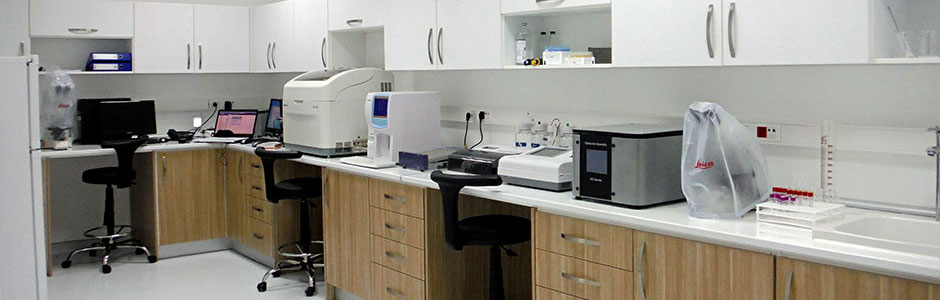
Laboratory Service