State-of-the-art radiographic, ultrasonographic and endoscopic devices are available in the Near East University Animal Hospital Video Diagnostics Unit. With these image techniques, images are taken from different parts of the patient after the first vet visit and the symptoms of the disease are tried to be discovered. For example, a cat or dog that has suffered a trauma, a traffic accident, or fallen from a height may have some bones fractures or lesions such as organ traumatization may have formed. In such a patient, X-rays of the skeletal system and films of the chest and abdomen are taken to see if there are any injured organs. If there are any suspicious findings in the films, the internal structure of the organs is examined by ultrasonographic examination, and it is checked whether there are any injuries or bleeding. Apart from this, if it is planned to raise newborn animals with a dog or cat, if the animal has started to give birth or has an internal disease and is not eating or drinking anything, if there is diarrhoea and vomiting, a diagnosis is made with the help of other findings by using these imaging techniques.
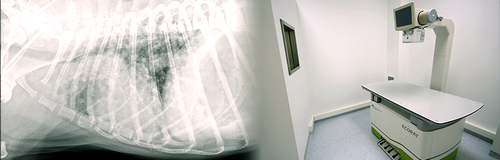
In the radiology unit, there is a digital radiography (DR) device that is specially designed for small animals and transfers images to digital media to serve this purpose. Thanks to this device, the images of the patient become visible on the screen in seconds, without the need for any other intervention in between. While evaluating these images created later, they can be enlarged as much as desired and examined by changing the brightness and contrast settings. These features provide great advantages over conventional radiography. With this device, patients of all sizes, from a chihuahua to a St. Bernhard breed dog, from a budgerigar to Ara parrots can be evaluated. After the images are taken, they can be saved, sent by e-mail, archived without deterioration, and thus, they are both useful for control examinations and an excellent source of material for educational and scientific studies.
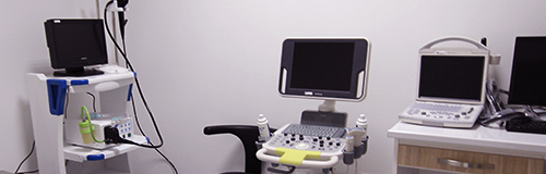
The ultrasonography device, which is mostly used in the examination of soft tissues, also has the Doppler function, which is one of the most advanced models of its kind, in which currents can be evaluated. Thanks to this function, heart, vascular and circulatory examinations can also be easily performed, thus helping with both internal diseases and childbirth. While ultrasonography transfers the cross-sectional image of the organs and tissues on which the probe is located, it also simultaneously shows the movements. Thus, without harming the patient, taking cell and tissue samples, such as fluid or biopsy, can be performed easily, or the babies and their heartbeats can be monitored in pregnant animals. These are just some small examples of the possibilities and the list could be expanded further.
For endoscopic examination, there is a bendable endoscope, and with the help of this instrument, the contents and inner surfaces of the organs can be examined by entering different body cavities. In this way, a diagnosis of a disease that does not heal easily, such as a foreign body that has escaped into the respiratory tract or been swallowed, or a stomach ulcer can be made. Here, the video is recorded and reflected on a large screen. In this way, both patient owners and students will have the opportunity to see the images again and get information and examine them. Again, it is possible to take another step towards the diagnosis of the disease by taking fluid and tissue samples with biopsy equipment under the guidance of endoscopy.